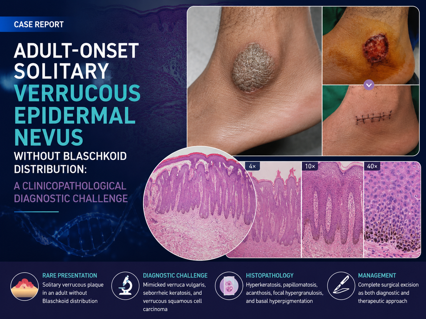
Adult-Onset Solitary Verrucous Epidermal Nevus Without Blaschkoid Distribution: A Clinicopathological Diagnostic Challenge
DOI:
https://doi.org/10.56127/jukeke.v5i2.2740Keywords:
verrucous epidermal nevus, Blaschko’s lines, histopathological examination, excisional biopsy, verrucous lesionAbstract
Verrucous epidermal nevus (VEN) is a benign keratinocytic hamartoma that usually appears in childhood and follows Blaschko’s lines. Adult-onset solitary VEN without Blaschko line distribution is rare and may clinically mimic other verrucous lesions, including verruca vulgaris, seborrheic keratosis, and verrucous squamous cell carcinoma. Objective: This case report aims to describe an atypical adult-onset solitary VEN without Blaschko line distribution and emphasize the importance of clinicopathological correlation in establishing the diagnosis. Case: A 27-year-old man presented with a progressively enlarging verrucous plaque on the left lateral malleolus for one year. The lesion was hyperkeratotic, well-demarcated, and associated with pruritus. Clinical differential diagnoses included verruca vulgaris, seborrheic keratosis, and verrucous squamous cell carcinoma. Complete surgical excision was performed, followed by histopathological examination. Discussion: Histopathological findings showed hyperkeratosis, papillomatosis, focal hypergranulosis, acanthosis, and basal hyperpigmentation without koilocytosis, horn cysts, cytologic atypia, or invasive growth. These findings confirmed the diagnosis of VEN and excluded infectious and malignant verrucous lesions. Conclusion: Solitary adult-onset VEN without Blaschko line distribution is a rare diagnostic challenge. Histopathological examination is essential for accurate diagnosis, while excisional biopsy may serve both diagnostic and therapeutic purposes in localized lesions.
References
Aghajani, M., Lu, J. T., Frew, J. W., & Sebaratnam, D. F. (2026). The evolving genetic landscape of ILVEN: A comprehensive review. Journal of the European Academy of Dermatology and Venereology, 40(3), 353-360. https://doi.org/10.1111/jdv.70098
Alaziz, M., & Najim, N. R. (2021). Histological differential diagnosis of verruca vulgaris: A systematic review. International Journal of Clinical and Diagnostic Pathology, 4(3), 26-31. https://doi.org/10.33545/pathol.2021.v4.i3a.385
Dağtaş, B. B., Manav, V., Leblebici, C., & Koku Aksu, A. E. (2025). Retrospective evaluation of 28 cases of inflammatory linear verrucous epidermal nevus. Turkish Archives of Dermatology and Venereology, 59(2), 54-59. https://doi.org/10.4274/turkderm.galenos.2025.44969
Gazali, M., Ruslin, M., Stevanie, C., Yusuf, A., Al-Jamaei, A. A., Boffano, P., Forouzanfar, T., & Tomihara, K. (2024). Short-term follow-up of surgical management verruca vulgaris with modified Estlander flap: A case report and recent literature review. Journal of Clinical and Experimental Dentistry, 16(1), e1033-e1039. https://doi.org/10.4317/jced.61814
Hafner, C. (2014). Mosaic RASopathies. Journal of Medical Genetics, 51(8), 473-474. https://doi.org/10.1136/jmedgenet-2014-102550
Hafner, C., Toll, A., Fernández-Casado, A., Earl, J., Marqués, M., Acquadro, F., Méndez-Pertuz, M., Urioste, M., Malats, N., Burns, J. E., & Real, F. X. (2012). Keratinocytic epidermal nevi are associated with mosaic RAS mutations. Journal of Medical Genetics, 49(4), 249-253. https://doi.org/10.1136/jmedgenet-2011-100637
Johnson, A. N., Sum, K., Rieger, K. E., Chiou, A. S., & Li, D. J. (2024). Adult-onset inflammatory linear verrucous epidermal nevus successfully treated with intralesional steroid. JAAD Case Reports, 46, 5-7. https://doi.org/10.1016/j.jdcr.2024.02.001
Mishra, K., Kant, S., & Verma, S. K. (2015). Verrucous epidermal nevus: A case report. Journal of Clinical and Diagnostic Research, 9(2), WD01-WD02. https://doi.org/10.7860/JCDR/2015/11082.5569
Montanari, A., Caroppo, F., Amabile, A., Fortina Belloni, A., & Bassetto, F. (2024). Suspect dyskeratotic neoformations in a 7-year-old child with keratitis-ichthyosis-deafness syndrome: Diagnostic, surgical and wound care management. Dermatology Reports, 16(4). https://doi.org/10.4081/dr.2024.9953
Morren, M. A., Fodstad, H., Brems, H., Bedoni, N., Guenova, E., Jacot-Guillarmod, M., Busiah, K., Giuliano, F., Gilliet, M., & Atallah, I. (2024). Mosaic RASopathies concept: Different skin lesions, same systemic manifestations? Journal of Medical Genetics, 61(5), 411-419. https://doi.org/10.1136/jmg-2023-109306
Nayak, S. U., Shenoi, S. D., Singh, R., & Pai, K. (2022). Adult-onset linear verrucous epidermal nevus in an Indian male. Clinical Dermatology Review, 6(2), 149. https://doi.org/10.4103/cdr.cdr_138_20
Verzì, A. E., Lacarrubba, F., Quattrocchi, E., & Micali, G. (2019). Verrucous epidermal nevus: Dermatoscopy, reflectance confocal microscopy and histopathological correlation. Dermatology Practical and Conceptual, 9(3), 230-231. https://doi.org/10.5826/dpc.0903a16
Waldman, A. R., Garzon, M. C., & Morel, K. D. (2022). Epidermal nevi. Dermatologic Clinics, 40(1), 61-71. https://doi.org/10.1016/j.det.2021.09.006
Ye, Q., Jia, M., Deng, L. J., & Fang, S. (2023). Cutaneous verrucous carcinomas: A review. Journal of Cutaneous Medicine and Surgery, 27(2), 150-156. https://doi.org/10.1177/12034754231155895
Zuntini, R., Cattani, C., Pedace, L., Miele, E., Caraffi, S. G., Gardini, S., Ficarelli, E., Pizzi, S., Radio, F. C., Barone, A., Piana, S., Bertolini, P., Corradi, D., Marinelli, M., Longo, C., Motolese, A., Zuffardi, O., Tartaglia, M., & Garavelli, L. (2023). Case report: Sequential postzygotic HRAS mutation and gains of the paternal chromosome 11 carrying the mutated allele in a patient with epidermal nevus and rhabdomyosarcoma: Evidence of a multiple-hit mechanism involving HRAS in oncogenic transformation. Frontiers in Genetics, 14, 1-10. https://doi.org/10.3389/fgene.2023.1231434